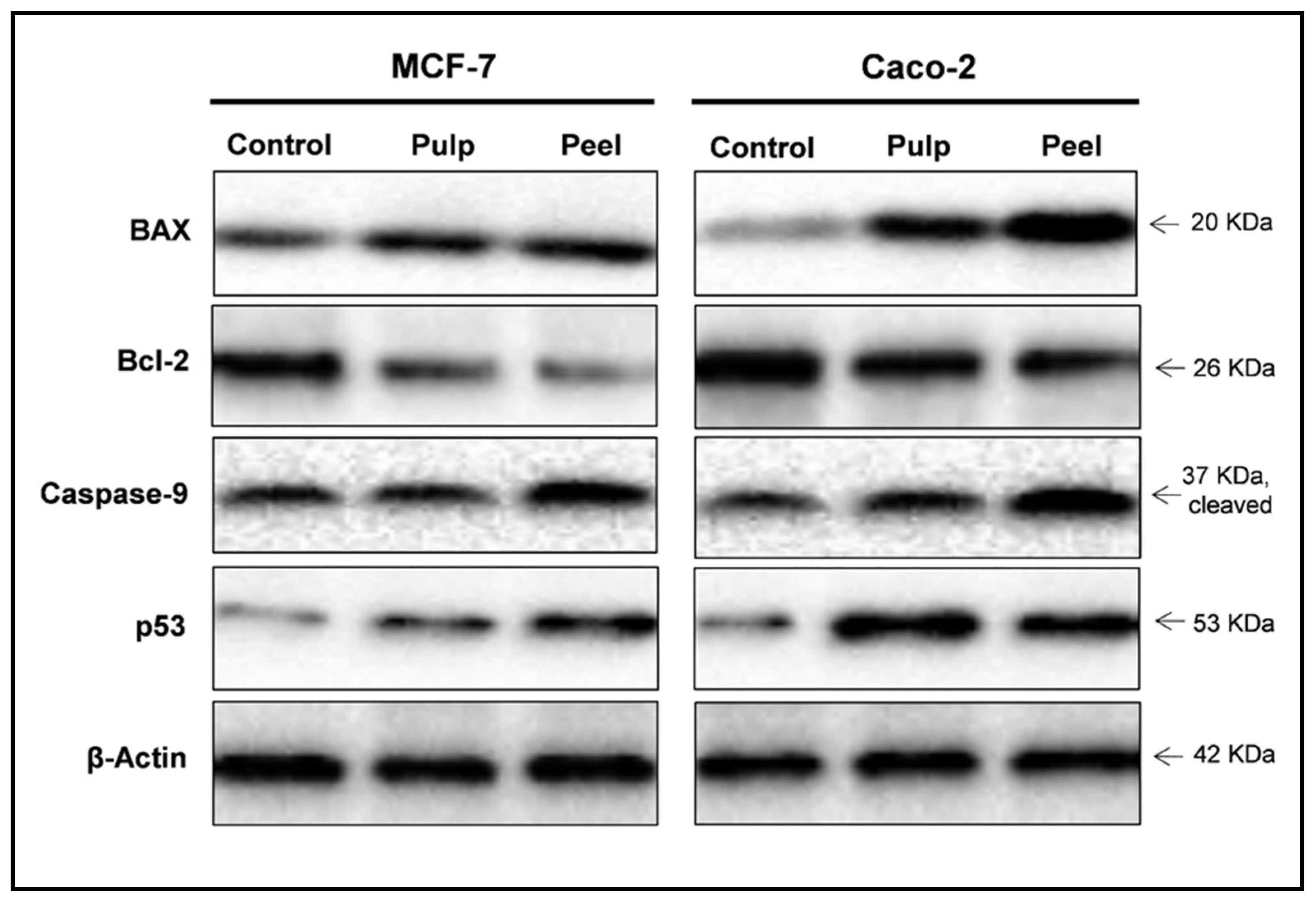
The indirect method offers many advantages over the direct method, which are described below. Labels (or conjugated molecules) may include biotin, fluorescent probes such as Invitrogen Alexa Flour or DyLight flourophores, and enzyme conjugates such as horseradish peroxidase (HRP) or alkaline phosphatase (AP). Subsequently, the primary antibody is detected using an enzyme- or fluororophore-conjugated secondary antibody. In the indirect detection method, an unlabeled primary antibody is first used to bind to the antigen.

This detection method is not widely used as most researchers prefer the indirect detection method for a variety of reasons. With the direct detection method, an enzyme- or fluorophore-conjugated primary antibody is used to detect the antigen of interest on the blot. One common variation involves direct versus indirect detection. Procedures vary widely for the detection step of a western blot experiment. Whatever system is used, the intensity of the signal should correlate with the abundance of the antigen on the membrane. Fluorescent blotting is a newer technique and is growing in popularity as it affords the potential to multiplex (detect multiple proteins on a single blot). Alternatively, fluorescently tagged antibodies can be used, which require detection using an instrument capable of capturing the fluorescent signal. However, digital imaging instruments based on charge-coupled device (CCD) cameras are becoming popular alternatives to film for capturing chemiluminescent signal. The light output can be captured using film. The most sensitive detection methods use a chemiluminescent substrate that produces light as a byproduct of the reaction with the enzyme conjugated to the antibody. Chromogenic substrates produce a precipitate on the membrane resulting in colorimetric changes visible to the eye. Often the secondary antibody is complexed with an enzyme, which when combined with an appropriate substrate, will produce a detectable signal. Most commonly, the transferred protein is then probed with a combination of antibodies: one antibody specific to the protein of interest (primary antibody) and another antibody specific to the host species of the primary antibody (secondary antibody). Next, the membrane is blocked to prevent any nonspecific binding of antibodies to the surface of the membrane. Subsequently, the separated molecules are transferred or blotted onto a second matrix, generally a nitrocellulose or polyvinylidene difluoride (PVDF) membrane. 3 March 2016.The first step in a western blotting procedure is to separate the macromolecules in a sample using gel electrophoresis. American Association for the Advancement of Science. Instructions for preparing an initial manuscript.American Society for Biochemistry and Molecular Biology. (2015) An analysis of critical factors for quantitative immunoblotting. (2015) Considerations when quantitating protein abundance by immunoblot. McDonough AA, Veiras LC, Minas JN, Ralph DL.Degasperi A, Birtwistle MR, Volinsky N, Rauch J, Kolch W, Kholodenko BN (2014) PLoS ONE.Get in TouchĬontinue reading: Using Chemistry to Get Proportional Signals LI-COR provides products, protocols, and support for Western blotting (and a range of other protein assays) that help reduce variability and increase confidence in your results. What steps can you take today to improve your Western blot results? How many technical and biological replicates do you need?.

How will the way you compare and analyze quantified bands affect your results?.What normalization strategy works best for your experimental context?.Does the amount of protein you are loading allow for accurate quantitation?.What is the dynamic range of your detection system?.Is your detection chemistry based on chemiluminescent or fluorescent detection?.Regardless of your level of expertise, there are multiple questions to answer before you begin. Maybe you’ve been doing Westerns for years. Maybe you’re about to attempt your first one.


 0 kommentar(er)
